Oracle IAS, the best coaching institute for UPSC/IAS/PCS preparation in Dehradun brings to you UKPCS Science Life Sciences (paper 6)-Human Ear: Structure and Functions
Introduction
Each human ear consists of three portions:
(i) External ear,
(ii) Middle ear and
(iii) Internal ear.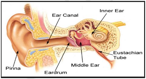
-
Human Ear- External Ear:
It comprises a pinna, external auditory meatus (canal) & tympanic membrane.
(i) Pinna:
The pinna is a projecting elastic cartilage covered with skin. Its most prominent outer ridge is called the helix. The lobule is the soft pliable part at its lower end composed of fibrous and adipose tissue richly supplied with blood capillaries. It is sensitive as well as effective in collecting sound waves.
(ii) External Auditory Meatus:
It is a tubular passage supported by cartilage in its exterior part and by bone in its inner part. The meatus (canal) is internally lined by hairy skin (stratified epithelium) and ceruminous glands (wax glands). The latter are modified sweat glands which secrete a waxy substance— the cerumen (ear wax) which prevents the foreign bodies entering the ear.
(iii) The tympanic membrane (tympanum):
Separates the tympanic cavity from the external auditory meatus. It is thin and semi-transparent, almost oval, though somewhat broader above than below. The central part of the tympanic membrane is called the umbo. The handle of the malleus is firmly attached to the membrane’s internal surface.
Functions of External Ear:
It directs sound waves towards the tympanic membrane. The sound waves produce pressure changes over the surface of the tympanic membrane. The cerumen (ear wax) prevents the entry of the foreign bodies into the ear.
-
Human Ear- Middle Ear:
It includes the following:
- The tympanic cavity, filled with air is connected with the nasopharynx through the Eustachian tube (auditory tube), which serves to equalize the air pressure in the tympanic cavity with that on the outside.
- There is a small flexible chain of three small bones called ear ossicles— the malleus (hammer shaped), the incus (anvil shaped) and the stapes (stirrup shaped).
The malleus is attached to the tympanic membrane on one side and to the incus on the other side.
The incus in turn is connected with the stapes, which is attached to the oval membrane covering the fenestra ovalis (oval window) of the inner ear. Malleus is the largest ossicle, however, stapes is smallest ossicle. Stapes is also the smallest bone in the body.
Functions of Middle ear:
(i) Due to the pressure changes produced by sound waves, the tympanic membrane vibrates, i.e., it moves in and out of the middle ear. Thus the tympanic membrane acts as a resonator that reproduces the vibration of sound,
(ii) It transmits sound waves from external to the internal ear through the chain of ear ossicles,
(iii) The intensity of sound waves is increased about twenty times by the ear ossicles. It may be noted that the frequency of sound does not change and from the tympanic cavity extra sound is carried to the pharynx through Eustachian tube.
3. Human Ear-Internal Ear:
There is a body cavity on each side enclosed in the hard periotic bone which contains the perilymph. The later corresponds to the cerebrospinal fluid. A structure, the membranous labyrinth floats in the perilymph. The membranous labyrinth consists of three>semicircular ducts, utricle, saccule, endolymphaticus and cochlea.
(i) Semicircular Ducts:
There are present three semicircular ducts; the anterior, the posterior and the lateral semicircular ducts.
Each semicircular duct is enlarged at one end to give rise to a small rounded ampulla.
Each ampulla contains a sensory patch of cells, the crista .The cristae are concerned with balance of the body.
(ii) Utricle, Endolymphaticus and Saccule:
The utricle is a dorsally placed structure to which all the three semicircular ducts are connected. The saccule is a ventrally situated structure which is joined with the utricle by a narrow utriculosaccular duct. From this duct a long tube, the ductus endolymphaticus arises which ends blindly as the saccusendolymphaticus.
(iii) Cochlea:
It is the main hearing organ which is connected with saccule by a short ductus reuniens leading from the saccule. It is spirally coiled that resembles a snail shell in appearance. It tapers from a broad base to an almost pointed apex.
Its properties are to determine the patterns of vibration of sound waves.
FUNCTION OF EAR
The ear performs the functions of hearing and balancing (equilibrium).
MECHANISM OF HEARING
The sound waves are collected by the external ear up to some extent. They pass through the external auditory meatus to the tympanic membrane which is caused to vibrate. The vibrations are transmitted across the middle ear by the malleus, incus and to the stapes bones.
From the perilymph the vibrations are transferred to the scala vestibuli of cochlea and then to scala media through Reissner’s membrane.
The impulses thus received by the hair cells are carried to the brain (temporal lobe of each cerebral hemisphere) through the auditory nerve where the sensation of hearing is felt (recognised).
Contact us for:-
-
- IAS coaching in Dehradun (Uttarakhand)
- UKPCS/UPPCS/UPPSC Mains coaching in Dehradun (Uttarakhand)
- Current Affairs classes in Dehradun (Uttarakhand)
- For getting detailed feedback on your answers and improve answer writing
- Phone Number:–9997453844

He is a bibliophile,accomplished writer and a fitness enthusiast.
- UKPCS Upper Prelims 2026: Full-Length Mock Test Series by Oracle IAS - July 18, 2026
- 91 Oracle IAS Students Selected in UKPCS 2024 Upper Exam - June 3, 2026
- UKPSC अपर प्रीलिम्स 2026 टेस्ट सीरीज़ || Oracle IAS - April 10, 2026